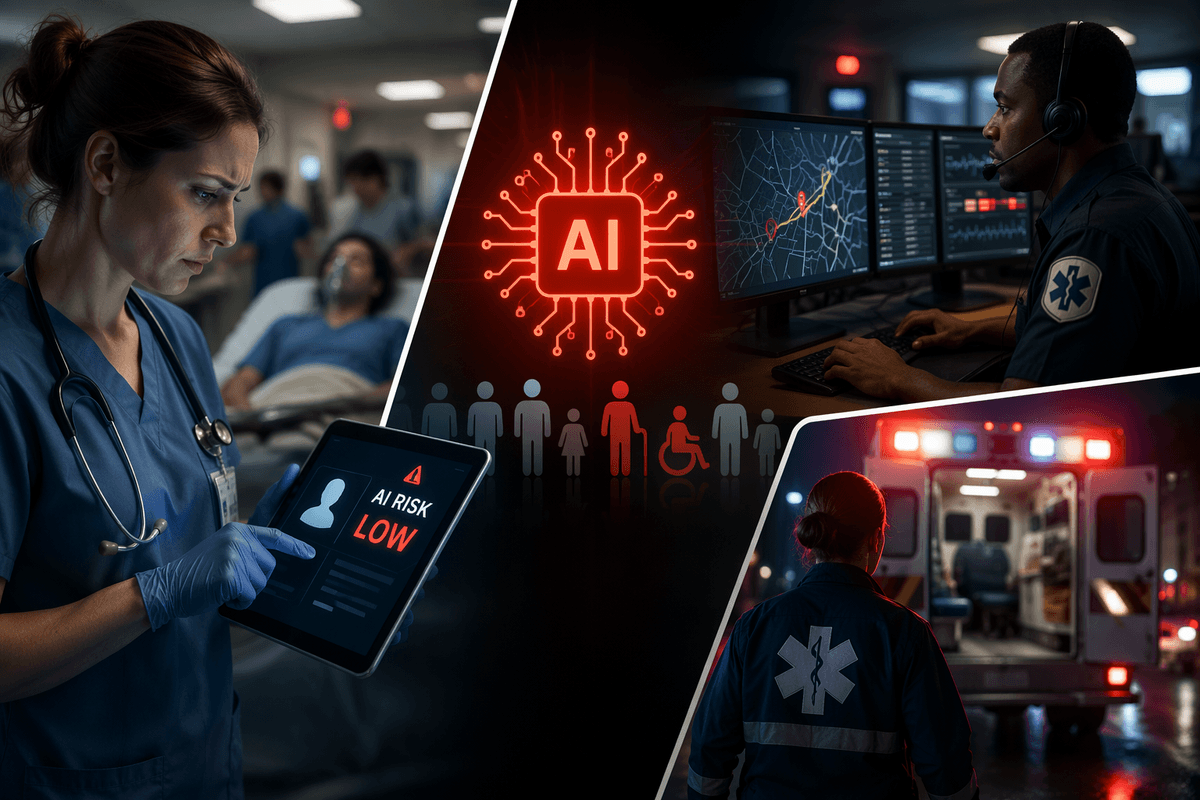
Why chest X-ray turnaround is still painfully slow
A chest X-ray can be taken in minutes, but the interpretation time is shaped by everything around the image. Studies queue behind CTs, trauma films, and inpatient follow-ups, and “stat” labels often outnumber true emergencies. Radiologists work within batching, shift coverage, and interruption-heavy environments where phone calls and protocol questions fragment attention. The image itself may be easy; the context rarely is—missing history, unclear indication, or no prior comparison can slow a read more than subtle findings.
The bottleneck is also logistical: getting the right exam linked to the right patient, resolving repeat or portable studies, and ensuring results reach the person who can act. Even when the report is signed quickly, downstream steps—alerting the team, reconciling incidental findings, documenting follow-up—add time. Speed improvements have to respect this reality, or they simply move delays to a different part of the chain.
Where AI helps most: triage, detection, and prioritization
Picture a worklist with dozens of chest X-rays that all look equally urgent on paper. AI helps most when it reshapes that queue: flagging exams more likely to contain time-sensitive findings (for example pneumothorax, large pleural effusion, new consolidation, or malpositioned lines/tubes) so a radiologist sees them sooner. That can reduce the time to action even if total reading volume stays the same.
Detection is typically narrow and pattern-based: it can highlight regions of interest, quantify change against priors, or surface “possible abnormal” cases that deserve an earlier look. The sensitivity comes with false alarms; if an algorithm over-flags, clinicians learn to ignore it and the worklist becomes noisy. The best use is prioritization support, not an automated final read, with responsibility still resting on the interpreting clinician.
Speed versus accuracy: what metrics actually matter
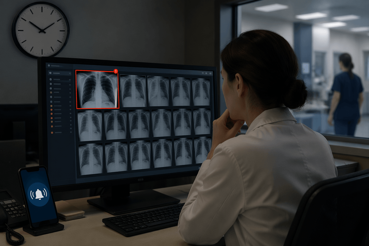
A common failure mode in evaluating AI for chest X-ray is treating “speed” as minutes saved per read. What matters more is time to the first appropriate action: time-to-notification for a critical finding, time until antibiotics are started for a suspected pneumonia, or time until a malpositioned line is corrected. An algorithm that shortens those intervals by moving the right studies to the top of the worklist can be clinically valuable even if overall report turnaround time barely changes.
Accuracy also needs to be tied to the use case. Sensitivity for “can’t miss” findings (like pneumothorax) may be prioritized over specificity, but only up to the point where false positives flood the queue and create alert fatigue. Ask for performance at clinically relevant thresholds, stratified by setting (ED vs ICU vs outpatient) and by prevalence, because positive predictive value will drop when the finding is rare. AUC looks good on a slide; action-oriented operating points are what change care.
How AI fits into radiology workflow without creating new work
In practice, workflow-friendly AI behaves like a quiet layer inside the tools people already use. The cleanest integrations place an indicator and a confidence score directly on the PACS/RIS worklist, optionally boosting certain studies upward based on agreed rules (for example, “possible pneumothorax” above routine follow-ups). Radiologists can then open the exam as usual, with an overlay available on demand rather than forced into the reading sequence. When the AI output requires extra clicks, separate logins, or manual exporting, it rarely saves time—it adds it.
Sites that avoid “new work” are explicit about what the model is allowed to trigger. Many limit automation to prioritization and a small set of critical alerts routed through existing channels, with clear labeling that AI is advisory. Someone must maintain thresholds, monitor drift, and handle downtime paths, or the system becomes either ignored noise or an unsafe shortcut.
Safety, bias, and accountability in real clinical conditions
A radiology department rarely experiences the “clean” conditions used to validate chest X-ray AI. Portable films, rotated patients, lines and tubes, low inspiration, and missing priors can all shift performance, and the errors are not random: a triage model that occasionally misses subtle pneumothorax in ICU portables is more consequential than one that overcalls mild atelectasis. Ask to see results by acquisition type (portable vs PA/lateral), care setting, and prevalence, because the same sensitivity can translate into very different false-alarm rates.
Bias shows up as uneven error across patient groups and sites: different equipment, body habitus, and comorbidity patterns can change what “normal” looks like. Accountability also has to be explicit. If AI reorders the worklist or triggers notifications, define who owns threshold changes, who reviews missed/overcalled cases, and what happens during downtime. In most deployments, the radiologist remains responsible for the final interpretation, so governance is a real, ongoing cost—not a one-time checkbox.
Buying or building: how to evaluate AI tools quickly
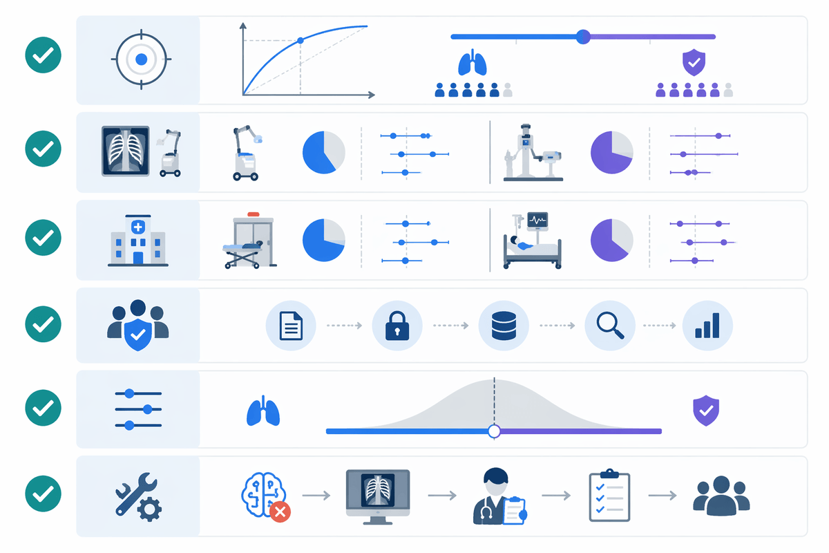
The fastest way to evaluate “buy vs build” is to start with the decisions you need the tool to support: worklist reprioritization, critical finding notification, or second-read highlighting. Most departments end up buying because building means labeled data pipelines, model maintenance, MLOps staffing, security reviews, and a plan for performance drift—costs that don’t disappear after go-live. Even when a model is technically strong, integration and governance usually dominate timelines.
For a quick but meaningful vendor screen, ask for evidence at a fixed operating point (your chosen sensitivity/specificity), plus results by portable vs PA/lateral and by ED vs ICU. Require a local “silent mode” test where AI runs without changing workflow, so you can measure false alarms, time-to-action impact, and failure modes on your equipment. Also confirm the downtime path, who can change thresholds, and how outputs are logged for audit and feedback.
Planning rollout: pilots, thresholds, and measurable outcomes
The first rollout decision is where the model is allowed to influence the queue. A practical pilot is a “silent” phase (AI scores logged but not shown), followed by a limited display phase (badge on the worklist, no auto-reordering), and only then rule-based reprioritization for a narrow set of findings. Pick thresholds with the people who will live with them: the radiologist team, the ED/ICU stakeholders, and whoever receives alerts. Too sensitive means constant interruptions; too strict means missed opportunities and loss of trust.
Define outcomes that are hard to game: time-to-notification for pneumothorax, time-to-action on line malposition, percent of critical findings acted on before report sign, and radiologist time spent managing AI outputs. Track false-positive alert rate per 100 studies, “AI down” frequency, and how often clinicians override prioritization rules, because those measures reveal whether speed is real or just rework.
What “faster” should look like after implementation
You notice “faster” first when the right cases surface earlier without extra clicking: ICU portables with a likely pneumothorax rise to the top, line malpositions get reviewed before the next med pass, and ED teams receive fewer-but-better critical notifications. Total report turnaround may improve modestly, but the meaningful win is shorter time-to-notification and time-to-action for a defined set of findings, tracked week over week.
Faster should not mean more interruptions or a louder worklist. If radiologists spend time adjudicating low-value flags, speed gains evaporate. A realistic target is a measurable drop in critical delays with a stable false-alert rate per 100 studies, plus a clear downtime plan so performance doesn’t depend on the AI being available.